Docket #: S13-195
3D printed smartphone lens adapters for mobile anterior and posterior segment ophthalmoscopy
Stanford researchers have designed and prototyped an inexpensive, compact and easy-to-use smartphone lens mount for the capture of high quality photographs and videos of the eye's front and back structures. Using standard lenses and this 3D printed adapter, it is possible to perform anterior segment and indirect ophthalmoscopy. This invention also eliminates the need for expensive and less portable equipment, such as a slit lamp or a Panoptic ophthalmoscope, and greatly simplifies manually aligning the lens, light source, and camera. When coupled with an appropriate mobile app and encryption, this user-friendly adapter requires little to no specialized training, such that patients as well as healthcare practitioners may acquire the images. To capitalize on this invention's ability to aid healthcare providers' and patients' rapid communication in both modern and developing worlds see the list of applications below.
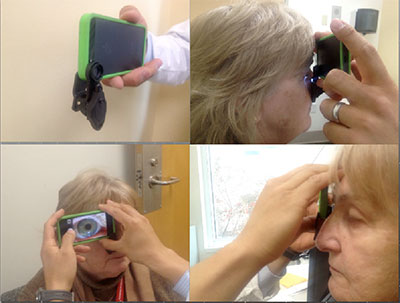

Caption: Ophthalmoscopy condensing lens mounted to an iPhone 5 and a fundus photo taken with the attachment.
Stage of Research
Prototyped 3 types of 3D printed lens adapters and preformed user testing. The results show the prototype can successfully enabling rapid acquisition of high-quality fundus photographs of patients in the clinic and emergency room setting.
Related Technologies: S15-099 is a continuation-in-part of this invention. It provides improvements over 13-195 by enabling increased user friendliness and universality of fit to phones, and accessibility by providing low cost lenses, as well as additional examination features including the possbility of both direct and indirect ophthalmoscopy.
Applications
- a simple, compact, easy-to-use attachment which enables smartphones to capture high quality photographs and videos of the structures of the front and the back of the eye by using an improved light source and lens setup
- optometry offices
- developing nations
- clinics
- underserved school districted
Advantages
- Low cost production -3D printing or injection molding (estimated adaptor cost is less than $100, which is far less expensive than slit lamps which cost thousands of dollars)
- User friendly – requires little to no specialized training
- Rapid image-capture
- Portable and durable
- Simple design
- Utilizes standard indirect ophthalmoscopy condensing lenses
- Compatible with current smartphones or tablets
- Slimline compact design
- Utilizes a separate adjustable light source and advanced software image processing to speed up and enhance image selection
- Easily detachable for convenient removal
Publications
- K.Y. Lai, et al Assessment of Eye Disease and Visual Impairment in the Nursing Home Population Using Mobile Health Technology Ophthalmic Surgery, Lasers and Imaging Retina 2020;51:262-270.
- S. Collon… Utility and Feasibility of Teleophthalmology Using a Smartphone-Based Ophthalmic Camera in Screening Camps in Nepal Asia-Pacific Journal of Ophthalmology Feb 2020.
- Myung, D., Jais, A., He, L., Blumenkranz, M. & Chang, R. (2014). 3D Printed Smartphone Indirect lens Adapter for Rapid, High Quality Retinal Imaging. Journal of Mobile Technology in Medicine, Volume 3:1 p. 9-15. Retrieved from http://www.journalmtm.com/2014/3d-printed-smartphone-indirect-lens-adapter-for-rapid-high-quality-retinal-imaging/
- Spector, R. (2014, March 7). Smartphones become 'eye-phones' with low-cost devices developed by ophthalmologists, Stanford News. Retrieved from http://med.stanford.edu/news/all-news/2014/03/smartphones-become-eye-phones-with-low-cost-devices-developed-by-ophthalmologists.html
Related Publications
M.W.M. Wintergerst, et al Diabetic retinopathy screening using smartphone-based fundus imaging in India Ophthalmology Article in Press 2020.
M.W.M. Wintergerst, et al Non-contact smartphone-based fundus imaging compared to conventional fundus imaging: a low-cost alternative for retinopathy of prematurity screening and documentation Nature Scientific Reports 2019.
Patents
- Published Application: WO2014194182
- Published Application: 20160113489
- Published Application: 20170280996
- Published Application: 20190269325
- Issued: 9,706,918 (USA)
- Issued: 10,092,182 (USA)
Similar Technologies
-
Multi-Functional In Vivo Cardiovascular Imaging Using Near-Infrared II Fluorescence S12-195Multi-Functional In Vivo Cardiovascular Imaging Using Near-Infrared II Fluorescence
-
Ultrasonic neuromodulation with Pattern Interference Radiation Force (PIRF) S16-382Ultrasonic neuromodulation with Pattern Interference Radiation Force (PIRF)
-
Method to visualize and quantify transitions in brain activity using topological data analysis S17-374Method to visualize and quantify transitions in brain activity using topological data analysis