Docket #: S14-321
Bright, cyan-excitable orange fluorescent proteins and bioluminescence reporters
Stanford researchers have engineered an exceptionally bright, cyan-excitable orange-red fluorescent protein (CyOFP) that can be used both for multiplex imaging with GFP and for high-sensitivity, bioluminescent in vivo imaging. This technology includes the isolated CyOFP and a CyOFP luciferase fusion protein that produces red photons (greater than 600nm) at 150x the rate of the firefly luciferase.
Because CyOFP can be induced by the same cyan light that induces GFP, it enables easy, simultaneous imaging of multiple structures or events without custom hardware. Furthermore, the CyOFP fusion protein provides bright photon output at red wavelengths that are effective for deep-tissue imaging in mammals, allowing sensitive detection of reporter proteins present in fewer than 1000 cells. This combination of features make CyOFP well-suited for advanced non-linear and light-sheet microscopy.
By overcoming in vivo sensitivity limits of bioluminescence, CyOFP could become the reporter of choice for tracking cell number, cell movement and gene expression in a small number of cells in live animals.
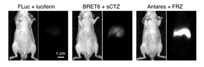
Bioluminescence of CyOFP fusion protein is superior to other reporters in living mice. The CyOFP fusion protein (Antares) with furimazine (FRZ) was injected into mice intravenously and compared with both BRET6 with coelenterazine (sCTZ) and firefly luciferase (FLuc2) with luciferin. Antares produced about 4x more detectable emission than BRET6 and about 18x more detectable emission than FLuc.
Stage of Development
The inventors have demonstrated that CyOFP enables single-excitation multiplexed imaging with GFP-based probes in single- and two-photon microscopy, including time-lapse imaging in light-sheet systems. They have also shown that the CyOFP fusion protein is a highly sensitive bioluminescent report in vivo, producing 18x more emission in mice than firefly luciferase.
Applications
- Advanced microscopy - enables red photon emissions simultaneous with GFP for multiplex imaging in cell-based or gene expression assays
- In vivo imaging:
- non-invasive, bioluminescent tracking of cell number, cell movement or gene expression in live mammals
- particularly useful for imaging small numbers of cells (1000) in deep tissue for stem cell implantation or preclinical testing of cancer therapeutics
- Bioluminescent resonance energy transfer (BRET) systems
Advantages
- Red photon emission (600nm):
- optimal for deep tissue imaging
- useful as companion reporter with well-established GFP reporters for detecting two structures or biological events
- Bright:
- CyOFP fusion protein produces red photons that transmit through mammalian tissue at 150x the rate of previous luciferases or BRET systems
- bright emission enables highly sensitive detection (1000 cells)
- Cyan-excitable:
- enables co-excitation of CyOFP and GFP using the same hardware in state-of-the art microscopy applications such as super-resolution imaging or advanced light-sheet and two-photon techniques
- faster results without any need to adjust for time shift from acquition of different image channels
Publications
- Chu, Jun, Younghee Oh, Alex Sens, Niloufar Ataie, Hod Dana, John J. Macklin, Tal Laviv et al. "A bright cyan-excitable orange fluorescent protein facilitates dual-emission microscopy and enhances bioluminescence imaging in vivo." Nature biotechnology 34, no. 7 (2016): 760.
Related Links
Patents
- Published Application: 20160376332
- Issued: 9,908,918 (USA)
Similar Technologies
-
Ultra Bright Lanthanide-Doped Nanoparticles for Luminescence Imaging S18-147Ultra Bright Lanthanide-Doped Nanoparticles for Luminescence Imaging
-
X-ray Molecular Imaging with Radioluminescent Nanoparticles S10-006X-ray Molecular Imaging with Radioluminescent Nanoparticles
-
Oral administration of fluorescent films for prolonged delivery of optical imaging agents S18-021Oral administration of fluorescent films for prolonged delivery of optical imaging agents