Docket #: S19-416
Creation of a flexible ultrasound system for real time acquisition of large fields of view
Stanford researchers at the Ferrara Lab have developed a volumetric ultrasound imaging that uses a motion controller to realize 3D imaging. This invention introduces a new transducer architecture with significantly improved image resolution. The prototype has been tested on phantom with plans for human trials.
Figure
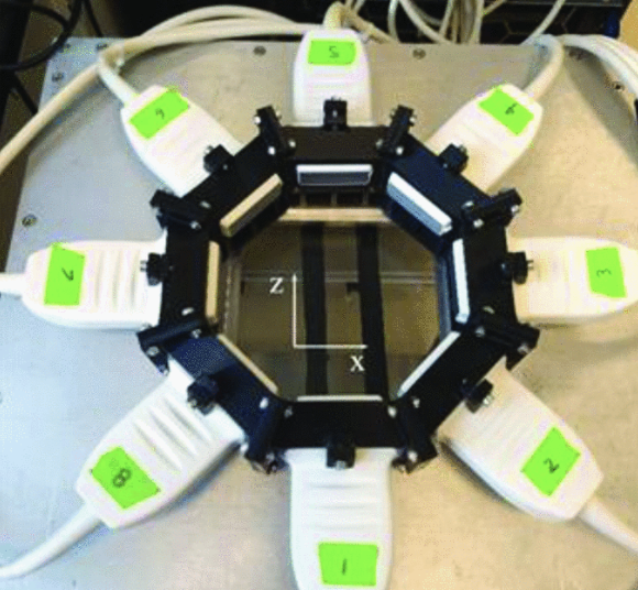
Figure description - The ring array assembled by eight L7-4 linear arrays and the orientation of the image grid.
In one embodiment, the invention uses a 1024-element matrix array transducer combined with 3D printed manifold to create arrays of calibrated and aligned transducers. A motorized system is incorporated to scan large regions of a subject under test, to enable use of software for 3D imaging and 3D passive acoustic mapping, and for combining imaging and therapy.
Image Credit-Publication below
Video:
Figure Description:
Realtime In vivo human imaging with the ultrasound tomography system. The volunteer moved the right hand and forearm up and down in the water tank.
Stage of Development
Applications
- 3D ultrasound monitoring and control
- Examples include but are not limited to:
- Microbubble cavitation for gene delivery for bone healing
- 3D imaging in pediatrics of the kidney, urogenital system, musculoskeletal system, hip, spine, liver
- 3D contrast agent imaging in adults
- Guiding ultrasound-based tomography
Advantages
- Significantly improved image resolution, contrast, contrast-to-noise ratio
- Customizable tailored geometry
- Can combine different types and different operating frequencies
- Excellent image quality
Publications
- Cai, Xiran, Josquin Foiret, Joseph Roth, Zulma Gazit, Gadi Pelled, Dan Gazit, and Katherine W. Ferrara. "3D monitoring and control of microbubble cavitation for gene delivery." In 2019 IEEE International Ultrasonics Symposium (IUS), pp. 888-890. IEEE, 2019.
Related Links
Similar Technologies
-
Ultrasonic neuromodulation with Pattern Interference Radiation Force (PIRF) S16-382Ultrasonic neuromodulation with Pattern Interference Radiation Force (PIRF)
-
(1) An improved method for operating CMUTs under high and varying pressure, and (2) Production of pre-charged CMUTs for zero-external-bias operation S11-214(1) An improved method for operating CMUTs under high and varying pressure, and (2) Production of pre-charged CMUTs for zero-external-bias operation
-
Phase-change nanoparticles for neuromodulation S17-163Phase-change nanoparticles for neuromodulation