Docket #: S16-093
Low Cost, High-Throughput CLARITY Imaging with Single-Cell Resolution and Axon Visualization
Researchers in Prof. Karl Deisseroth's laboratory have developed a low-cost, high-throughput system for high resolution light-sheet imaging to visualize individual cells throughout a volume of intact tissue, including directly visualizing and quantifying the axon tracts of neuronal circuits. This technology enables analysis of large sample cohorts using the CLARITY platform (a process which renders tissue transparent while leaving its cellular structures intact) by accelerating the experimental pipeline and reducing costs by over 90%. In addition, the new process is fully compatible with commercial platforms for imaging and data analysis, further reducing costs and complexity compared to the conventional CLARITY method. When the platform is used for mapping axon projection at micron-level resolution in intact brains, it enables both quantitative analysis of size and intensity/activity as well as direct visualization of region-to-region connectivity. This technology could be used to characterize tissue samples for diagnostics, to perform neuronal circuit analysis for basic neuroscience studies, or to identify cellular targets for therapeutic interventions.
Figure
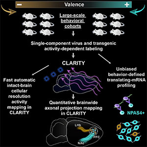
Schematic of high-throughput CLARITY work flow with axonal projection mapping as used for large scale analysis of prefrontal cortex wiring and associated behavior in mice.
Stage of Research
The inventors have demonstrated the utility of this technology by combining the high-throughput CLARITY pipeline with the axonal projection mapping to analyze the wiring of the prefrontal cortex in mice. These studies illuminated the logic of information processing in the brain by linking activity during behavior with the molecular properties of neurons.
Intellectual Property
This technology is described in two distinct patent applications, one for the high-throughput imaging system (Stanford Docket S16-092) and one for axonal visualization and quantification (Stanford Docket S16-093).
Applications
- Research and Therapeutic - high-throughput cellular imaging of intact tissue for activity and anatomical circuit mapping for basic neuroscience studies or to identify cellular targets for drugs or other therapeutic interventions
- Diagnostics - high-throughput imaging of intact samples to characterize tumors and other disease tissue
Advantages
- Fast, high-throughput analysis:
- accelerated, parallelized clarification enables analysis of large sample cohorts
- rapid, non-disruptive analysis of intact tissue (no sectioning)
- imaging under a single field of view and as a single stack in less than 2 hours
- Low cost - refractive index-matching process reduces costs by >90%
- High resolution:
- single-cell resolution throughout whole volume of tissue
- micron-level resolution of brain-wide axon tracts for quantitative analysis of fiber diameter and intensity/activity
- direct visualization of region-to-region physical connectivity
- Standard equipment - fully compatible with commercial platforms for imaging and data processing
- imaging with commercial light-sheet microscope
- data files from each tissue sample are small enough (~12GB) to be easily stored and directly analyzed on standard desktop work stations without compression or stitching
- no specialized clearing equipment such as electrophoresis or perfusion chambers
Publications
- Ye, Li, William E. Allen, Kimberly R. Thompson, Qiyuan Tian, Brian Hsueh, Charu Ramakrishnan, Ai-Chi Wang et al. "Wiring and Molecular Features of Prefrontal Ensembles Representing Distinct Experiences." Cell (2016).
- Stanford research shows that different brain cells process positive and negative experiences, Stanford Report May 26, 2016.
- PCT Published Patent Application WO2017205531A1, "Methods for visualization and quantification of fiber-like structures".
Related Links
Patents
- Published Application: WO2017205531
- Published Application: 20190187161
- Issued: 10,641,782 (USA)
Similar Technologies
-
Method and system for whole brain imaging and analysis S16-092Method and system for whole brain imaging and analysis
-
Light sheet fluorescence microscopy using high speed structured and pivoting illumination S16-332Light sheet fluorescence microscopy using high speed structured and pivoting illumination
-
Method for Fabrication of Arrayed Dual Axis Microscopes S13-036Method for Fabrication of Arrayed Dual Axis Microscopes