Docket #: S13-113
Super Resolution for Light Field Microscopy
Light field microscopy (LFM) is a new technique for high-speed volumetric imaging of weakly scattering or fluorescent specimens. It employs an array of microlenses to trade off spatial resolution against angular resolution, thereby yielding the information needed to reconstruct a volume from a single photographic exposure. However, this ability to perform scan-less 3-D imaging comes at a cost: the resulting volume reconstruction has considerably lower lateral resolution than a conventional microscope image.
This invention addresses this drawback of conventional light field microscopy. This technique, which draws its inspiration from“super-resolution” methods in computer vision, enables reconstructions of up to 8x higher resolution than previously possible with conventional LFM when reconstructing a planar object, and up to 2-4x higher resolution when reconstructing a sample with complex 3-D structure. This resolution improvement is due in part to a new, more accurate optical model based on wave optics that captures the effects of diffraction in light field microscope images. The GPU accelerated reconstruction algorithm in this implementation also performs 3-D deconvolution, which enhances lateral resolution while also computationally removing out-of-focus light in the volume for better optical sectioning.
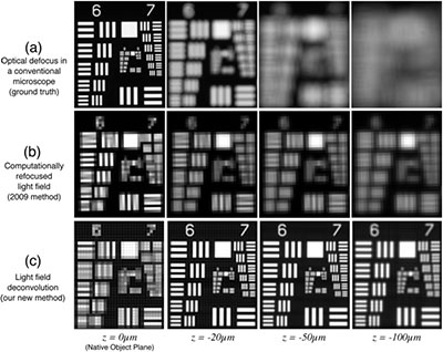
Comparison of conventional (a) and LFM (b and c) imaging of USAF 1951 resolution test target translated to depths up to 100 um from the native object plane (z = 0 um). The wave optics reconstruction algorithm (c) improves lateral resolution up to 8-fold compared to standard LFM imaging (b), except at the z = 0 plane (left image).
Stage of Research
The inventors have validated this technology by measuring lateral resolution on a standard USAF 1951 resolution target. They have demonstrating the ability to resolve higher spatial frequencies in the images reconstructed with the new algorithm with the target placed at a range of different z-depths spanning a range of +/- 200 microns. The technique has been shown to work well with a variety of different microscope objectives. They have also reconstructed images of pollen grain to demonstrate the improved image resolution and optical sectioning capability of a biological specimen.
Applications
- Microscopy - Light Field Microscopy for fast 3-D recording of dynamic phenomenon with end user applications in:
- biological research
- clinical pathology
- quality assurance inspections
Advantages
- High resolution 3-D imaging - 8-fold improved lateral resolution and better optical sectioning compared to standard LFM
- Improved Optical Model - the newly developed wave optics model is considerably more accurate when modeling the imaging process at microscopic scales where diffraction plays a key role.
Publications
- "Wave Optics Theory and 3-D Deconvolution for the Light Field Microscope, Michael Broxton, Logan Grosenick, Samuel Yang, Noy Cohen, Aaron Andalman, Karl Deisseroth, Marc Levoy, Optics Express, Vol. 21, Issue 21, pp. 25418-25439 (2013).
Patents
- Published Application: 20140263963
- Issued: 9,658,443 (USA)
Similar Technologies
-
Multimodal DAC Microendoscope Platforms S10-278Multimodal DAC Microendoscope Platforms
-
Robotic Microscopy System for Simultaneously Imaging Multiple Areas of a Sample S13-155Robotic Microscopy System for Simultaneously Imaging Multiple Areas of a Sample
-
Efficient wide-field nanosecond imaging methods using Pockels cells for low-light applications S18-388Efficient wide-field nanosecond imaging methods using Pockels cells for low-light applications