Docket #: S13-492
3D Super Resolution Microscopy with a Bisected Phase Mask: A Device to Measure the Depth, Position and Orientation of Single Molecules
Stanford researchers have designed a tunable wedge-based phase mask for 3D super-resolution imaging that can simultaneously determine both the position and rotational mobility of individual light-emitting molecules from a single camera image. This mask, termed the “bisected pupil” can be easily installed to augment commercial microscopes. The bisected pupil design includes a tunability parameter that may be optimized to precisely localize objects up to two microns from the objective lens focal plane. This enables the user to track the location and rotational behavior of multiple objects at different depths. The primary end-user application of this technology is for visualizing single fluorescently labeled molecules of interest in biological research. However, the technique is compatible with a variety of light-emitting labels (e.g. quantum dots, gold beads, or nanorods). A U.S. patent for this invention has been issued in November of 2017.
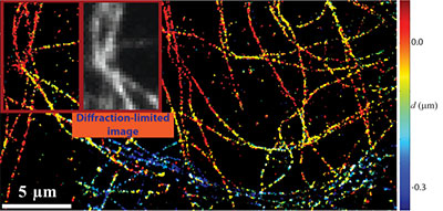
3D-super-resolved image of microtubules immunolabeled with Alexa-647 in a BSC-1 cell (Adapted from reference 1)
This technology modifies the point spread function of a conventional microscope so that fluorescent objects, such as single molecules, will appear as two 'spots' or 'lobes' on a conventional camera sensor. The depth of an object is encoded into the separation distance between the two lobes. Rotational mobility is assessed by measuring the relative brightness of the two lobes. We term this parameter the lobe asymmetry (LA) of a given object. When the lobes are of similar brightness (i.e. the lobe asymmetry is low), this indicates that an object is highly rotationally mobile. Additionally, using dual-polarization acquisition, another parameter, termed the linear dichroism (LD), may be simultaneously measured for each object, providing further information about rotational dynamics.


(a) The bisected pupil is implemented using a wedge-based phase mask. When using dual-polariztion acquisition, the blue and red arrows indicate the axis of polarization relative to the phase mask to be used for each of the two polarization channels (T and R indicate the polarization reflected and transmitted by a polarizing beamsplitter). (b) Simulated images of a single molecule as a function of depth. Note that the images in the two polarization channels appear rotated due to the particular geometry of our dual-polarization experimental setup. (c) Experimental calibration data of a fluorescent bead, used to relate lobe separation to depth. (Adapted from reference 1)

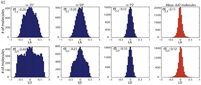
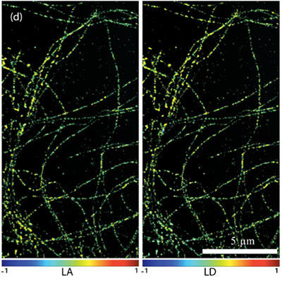
(a) Using a theoretical model for the rotational mobility of single molecules, experimental data was compared to simulation. (b) Image of a single DCDHF-N6 molecule in two polarization channels. The parameters LA and LD are computed from the raw intensities measured from the individual pixels of an acquired image. (c) By measuring the LA and LD of thousands to millions of fluorescent molecules, histograms are constructed, and compared to histograms generated from simulations. By comparing simulation to experimental data, we determine that the rotational mobility within a microtubule sample is very high. (d) By color-coding an image of microtubules according to the LA and LD of individual molecules, we conclude that the rotational mobility is uniform throughout the sample of interest. (Adapted from reference 2)
STAGE OF RESEARCH
The inventors used the BSP-modulated point-spread function to perform 3D super-resolved reconstructions of fluorescently labeled microtubules in fixed cells and simultaneously demonstrated that the rotational mobilities of the individual fluorescent dye molecules is great enough for accurate spatial localization. They have implemented a phase mask using a liquid crystal spatial light modulator (for rapid evaluation purposes), and have recently constructed a second-generation prototype using a custom-fabricated glass phase mask.

Photograph of a glass BSP phase mask (central optic). Fluorescence emission is passed through two relay lenses, and projected onto an EMCCD camera (far right) in order to form images with a modulated point spread function.
Applications
- 3D Microscopy with end-user applications such as:
- rapidly imaging thousands to millions of individual fluorescent molecules
- tracking precise 3D location of fluorescent or highly scattering particles
- characterizing rotational mobility of anisotropic particles
Advantages
- High resolution - 3D imaging of biological structures with resolution surpassing the diffraction limit by an order of magnitude
- Simultaneously measures depth and rotational motion of particles under observation
- Tunable extended depth of field (EDOF) parameter – for optimal imaging performance for objects up to 2 microns from focal plane
- Simple instrumentation :
- module with phase mask can be installed on a commercial microscope in less than 30 minutes
- rotational mobility data provided without additional cumbersome instruments
Publications
- Alex von Diezmann, Yoav Shechtman, W. E. Moerner, "Three-dimensional localization of single molecules for super-resolution imaging and single-particle tracking", Chem. Rev.. Vol. 117 (11), 7244-7275, (2017).
- A. S. Backer, M. P. Backlund, A. R. von Diezmann, S. J. Sahl, and W. E. Moerner, “A bisected pupil for studying single-molecule orientational dynamics and its application to 3D super-resolution microscopy,” Appl. Phys. Lett. 104, 193701 (2014).
- A. S. Backer and W. E. Moerner "Extending Single-Molecule Microscopy Using Optical Fourier Processing," J. Phys. Chem. B, Vol. 118 (28) 8313-8329 (2014).
Similar Technologies
-
Method for Fabrication of Arrayed Dual Axis Microscopes S13-036Method for Fabrication of Arrayed Dual Axis Microscopes
-
3D Super-Resolution Microscopy with Corkscrew Point Spread Function S10-1623D Super-Resolution Microscopy with Corkscrew Point Spread Function
-
Scanning microscope with very large field of view S13-154Scanning microscope with very large field of view