Docket #: S18-315
Airway visualization system
Stanford researchers designed a system to enable x-ray visualization of the tracheobronchial tree to aid the physician in guiding endoscopic tools in the pulmonary tract. Early diagnosis and treatment are vital for improving lung cancer survival rates, and tissue biopsy is necessary for diagnosis. However, performing a lung biopsy can be difficult because of the complexity of the highly branched airways of the lung, making it difficult to access and obtain the tissue and the procedure can pose a risk to the patient. Electromagnetic navigation bronchoscopy (ENB) is the safest way to obtain a biopsy but its diagnostic success is limited by poor real time visualization of significant portions of the lung. A major drawback to this method is the difficulty to visualize the lung airways in real-time in order to guide a bronchoscope. Should the biopsy procedure be unsuccessful, patients may then need to have the tissue surgically removed. In a high number of these cases, the nodules are found to be benign. This system will greatly improve the ability to obtain a diagnosis for the patient using ENB.
Stage of Research:
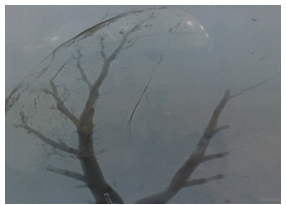
Ex vivo proof of concept showing detailed bronchogram in a pig lung
Applications
- Lung imaging to visualize the airways under fluoroscopic x-ray;
- Guided navigation during bronchoscopic guided lung nodule biopsy and treatment procedures; and
- Diagnosis of peripheral airway diseases such as asthma and COPD and infectious processes such as pneumonia or atelectasis.
Advantages
- Correct bronchoscopic positioning;
- More accurate navigation in the lung; and
- Higher diagnostic success rate for pulmonary nodule biopsies using ENB.
Publications
Patents
- Published Application: WO2019204499
- Published Application: 20200375448
- Issued: 12,004,707 (USA)
Similar Technologies
-
MagSweeper: high purity capture of circulating tumor cells and other rare cells S07-132MagSweeper: high purity capture of circulating tumor cells and other rare cells
-
Early diagnosis and treatment of eye cancer S19-500Early diagnosis and treatment of eye cancer
-
Automated single cell expression profiling in intact tissue by highly multiplexed in situ hybridization S16-313Automated single cell expression profiling in intact tissue by highly multiplexed in situ hybridization