Docket #: S11-248
The device and method to guide endothelial cell assembly
Stanford researchers have created a device with defined parallel-oriented fibrillar nanostructure that can control endothelial cell alignment along the direction of the fibrillar nanostructure. This device comprises collagen matrices and threads manufactured from solutions of clinical grade monomeric collagen in fibrillar form with regularly sized aligned fibrils, crimps, and angular distribution. The device and the process of orienting endothelial cells on the device mimic the effect of shear-induced cellular alignment in the ability to control cell morphology as well as cell function. Applications for this invention include bypass graft, AV shunt, implantable device, and bilayered graft.
On-going research:
Continuing to investigate the role of aligned collagen matrix proteins in modulating endothelial morphology and function.
Figure:
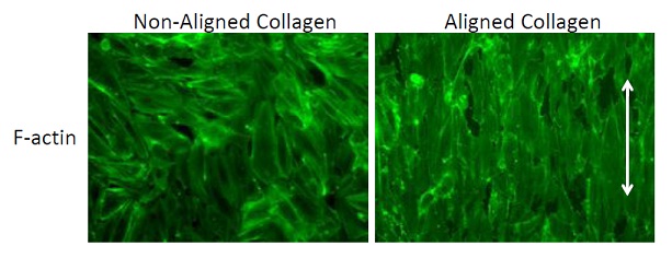
Figure Description: Endothelial cell morphology on aligned nanofibrillar collagen. Immunofluorescence staining of phalloidin (green) staining of cytoskeletal protein F-actin. Arrow denotes direction of collagen fibril assembly.
Applications
- Bypass graft- Endothelial cells at sites of anastamoses of bypass grafts are generally not aligned, which can promote the adhesion of lipogenic proteins or monocytes and thus lead to occlusions within the graft. By restricting the alignment of endothelial cells using fibrillar collagen matrix, the endothelial cells may be less prone to monocyte adhesion and may have improved patency.
- AV shunt
- Implantable device - Endothelial cells implanted to induce/stimulate/improve angiogenesis at the sites of compromised circulation, when delivered in a suspension format, usually do not survive long enough to exert any beneficial effects. Delivery of endothelial cells on fibrillar collagen thread improves their survival and may improve their angiogenic potential.
- Bilayered graft. Endothelial cells lining the interior wall of blood vessel are aligned along the vessel, while the smooth muscle cells comprising the outer layer of the vessel are helically aligned around the vessel axis. A bilayered graft with interior layer having the fibril alignment along the vessel axis, and the outer layer having a helical fibril alignment will provide guidance for endothelial cells to align inside the graft along its axis and for the smooth muscle cells on the outer surface of the graft to align in a helical orientation to the vessel axis. Aligning both endothelial and muscle layers according to their natural topography may improve patency and hoop strength of the graft. This model of the vessel may also be important for in-vitro study (e.g. drug discovery, device testing, etc.).
Advantages
- Mimics the effect of shear-induced cellular alignment in the ability to control cell morphology as well as cell function.
- Aligned collagen matrices reduce monocyte adhesion.
Publications
- Huang NF, Zaitseva T, Paukshto M, Sun J, Fuller G, Cooke JP. Vascular Cellular Morphology on Aligned Collagen Matrices. Biomedical Engineering Society Annual Conference, Austin, TX, Oct 6-9, 2010.
Related Links
Patents
- Published Application: WO2013103423
- Published Application: 20140242347
- Issued: 10,238,769 (USA)
Similar Technologies
-
Wireless, Ultra-Low Power Implantable Device S13-173Wireless, Ultra-Low Power Implantable Device
-
Magnetic Robotic Surgery Solution for Minimally Invasive Endovascular Procedures S23-047Magnetic Robotic Surgery Solution for Minimally Invasive Endovascular Procedures
-
Neuro-navigation device for localization of internal anatomical regions S17-412Neuro-navigation device for localization of internal anatomical regions