Docket #: S22-022
Modular PET scanning system that can be integrated into a MRI system
Stanford researchers have designed a modular PET scanning system that can be integrated into a MRI system. The proposed removable PET insert geometry enables a stand-alone MRI system to achieve large-field-of-view scanning, multi-bed body PET+MR studies appropriate for imaging any part of the body (e.g. the thorax) with large axial coverage.
In a conventional combined PET + MRI system, the two systems are permanently integrated together. This invention is lower cost and more flexible since it can be inserted into an existing stand-alone MRI system. However, current PET inserts to achieve PET+MRI have limited scanning capacity (e.g. head only). A key advantage of this invention is its ability to scan any part of the body.
Stage of Development
Figure:
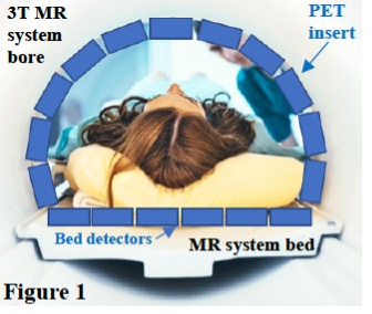
Description – PET insert
Image credit - Molecular Imaging Instrumentation Lab (MIIL)
Applications
- Dual modality – PET and MRI imaging
Advantages
- Modular – can be integrating into existing MRI systems
- Lower cost:
- Does not require use of expensive cryogens like helium-3
- Does not require the use of magnetic fields (distinct from adiabatic demagnetization technologies)
- Simplified process
- Technical advantages such as minimal stray magnetic field
Related Links
Similar Technologies
-
A noise robust decoder for multiplexing readout channels on an imaging sensor array S10-178A noise robust decoder for multiplexing readout channels on an imaging sensor array
-
Deep Learning Enabled Hybrid CT-MRI with Highly Sparse Sensory Data S21-073Deep Learning Enabled Hybrid CT-MRI with Highly Sparse Sensory Data
-
Optically coupled readout front-end for an MR-compatible PET system S07-046Optically coupled readout front-end for an MR-compatible PET system