Docket #: S24-145
Novel Radiopharmaceutical for Non-Invasive Imaging of Ferroptosis in Cancer Therapy
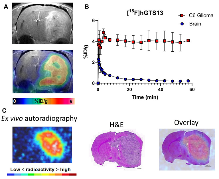
Stanford researchers in Prof. Corinne Beinat's lab have developed a novel radiotracer, [18F]hGTS13, for non-invasive imaging of system xc- activity, enabling the identification of ferroptosis-sensitive cancers and monitoring the efficacy of ferroptosis-inducing therapies. This technology facilitates personalized cancer treatment by assessing drug engagement and predicting therapeutic outcomes.
Ferroptosis, a form of regulated cell death driven by the iron-dependent accumulation of membrane lipid peroxides, has emerged as a promising target for cancer therapy, particularly in glioblastoma multiforme (GBM). However, the current state of the field lacks non-invasive imaging techniques to monitor the engagement of ferroptosis-inducing drugs and to identify patients who would benefit from such therapies. Existing radiotracers, like [18F]FSPG, have limitations in specificity and uptake in inflammatory cells, leading to suboptimal cancer imaging.
The novel radiotracer [18F]hGTS13 specifically targets system xc- to enable non-invasive imaging of ferroptosis in cancer cells. It offers improved radiosynthesis, enhanced specificity for cancer cells over inflammatory cells, and a high tumor-to-brain ratio in glioma models, making it a superior tool for personalized cancer treatment. Evidence from preclinical studies in rats demonstrates its effectiveness in distinguishing ferroptosis-sensitive and resistant cell lines and monitoring drug engagement, highlighting its ability to monitor drug engagement and efficacy in vivo, advancing the current state of cancer therapy.
Figure
Stage of Development
Proof of concept - in vivo data in a rat model of glioma
Related Technology
Docket S24-289: Theranostic for Targeted Treatment of Cancers
Applications
- Novel Imaging Agent: For Positron Emission Tomography (PET) and Computerized Tomography (CT)
- Cancer Therapy Monitoring: Non-invasive imaging to assess the efficacy of ferroptosis-inducing drugs in cancer treatment
- Drug Engagement Assessment: Monitors in vivo engagement of system xc- inhibitors, aiding in therapeutic development and optimization
Advantages
- First-In-Kind radiotracer to monitor cancer ferroptosis
- Broad Utility to monitor various cancers such as primary brain cancers, brain metastases, breast cancer, lung cancer, pancreatic cancer, liver cancer, lymphoma, head and neck cancer, ovarian cancer, or prostate cancer
- Enhanced Specificity: [18F]hGTS13 shows reduced uptake in inflammatory cells, improving cancer specificity over [18F]FSPG
- Improved Radiosynthesis: Incorporation of a UV-active group facilitates easier radiosynthesis and quality control compared to existing radiotracers
Publications
- Moses, A., Malek, R., et al. (2025). Monitoring of cancer ferroptosis with [18F] hGTS13, a system xc-specific radiotracer. Theranostics, 15(3), 836.
Related Links
Similar Technologies
-
Theranostic for Targeted Treatment of Cancers S24-289Theranostic for Targeted Treatment of Cancers
-
Cell-Penetrating, Guanidinium-Rich Oligomers for Drug and Probe Delivery S15-370Cell-Penetrating, Guanidinium-Rich Oligomers for Drug and Probe Delivery
-
Oligocarbonate molecular transporters across biological barriers S08-441Oligocarbonate molecular transporters across biological barriers