Docket #: S13-155
Robotic Microscopy System for Simultaneously Imaging Multiple Areas of a Sample
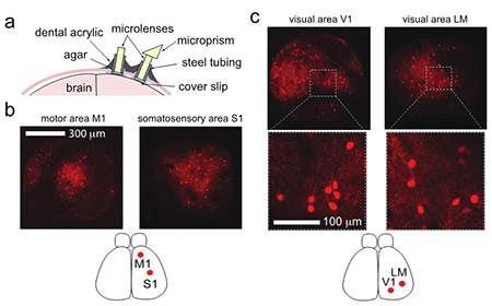
Researchers in Prof. Mark Schnitzer's laboratory have developed a robotic optical microscopy system which enables users to simultaneously view and record separate areas of a single three-dimensional sample. This system uses miniature objectives mounted on robotic arms to maneuver around a sample with five degrees of freedom. This allows cellular-level imaging in multiple places to visualize long-range interactions of cells distributed around the sample. This technology was designed for functional studies of neuron populations throughout the brain and could be used for other basic biomedical research or surgical robotics.
Figure
Stage of Development
Prototype
Applications
- In vivo imaging
- Simultaneous distal region imaging
- Neuroscience research
- Coupling with optical implant systems
- Robotic surgery
Advantages
- Omnidirectional sample access
- High speed (50 kHz camera)
- Customizable with single or multiphoton imaging
- Cellular resolution
Related Technology
Stanford Docket S24-107: Robotic Microscopy Platform for Imaging and Manipulating Biological Samples
Related Links
Patents
- Published Application: 20150057550
- Issued: 9,398,935 (USA)
Similar Technologies
-
Scanning microscope with very large field of view S13-154Scanning microscope with very large field of view
-
Breakthrough Optical Frequency Processing for Quantum Computing and Beyond S24-365Breakthrough Optical Frequency Processing for Quantum Computing and Beyond
-
Multimodal DAC Microendoscope Platforms S10-278Multimodal DAC Microendoscope Platforms