Docket #: S21-210
Temporally multiplexed one-photon and two-photon microscopy for neuroscience and spatial biological imaging
Stanford researchers have developed a device that combines one-photon and two-photon microscopy using fast temporal multiplexing enabling 3D alignment between in vivo and ex vivo data for neuroscience and spatial biology applications.
High-resolution, three-dimensional fluorescence imaging promises rich information about biological function and structure with applications ranging from neuroscience to oncology to spatial genomics. While nonlinear multiphoton microscopy provides the resolution and depth information to produce 3D images of tissues at single-cell resolution, but it has a limited field of view, slow acquisition times, and must be performed in a controlled laboratory setting not compatible with many in vivo and functional imaging studies. On the other hand, one-photon fluorescence imaging can be used for in vivo imaging due to its portability and large field of view, but it produces a low resolution, low depth-of-field image only.
The invented device aligns one-photon imaging with multi-photon ex vivo images at the cellular level to obtain three-dimensional data at single-cell resolution during in vivo functional studies. The inventors demonstrate its utility in neuroscience applications by imaging functional neural ensembles in mice to determine neuron identity, the morphology of neurons, and fidelity of the time traces of genetically encoded calcium indicators expressed in these neurons. This technology will find use in functional neuroscience imaging such as this but also has applications in spatial biology, including spatial proteomics and transcriptomics.
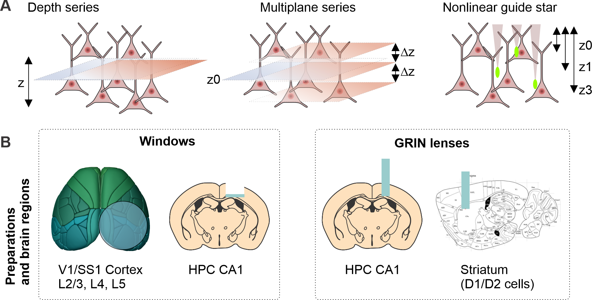
Figure (provided by inventors): Neuronal imaging in live mice with this technology
Applications
- Alignment of widefield functional in vivo imaging and three-dimensional, single-cell resolution ex vivo imaging with applications in:
- Functional neuroscience studies
- Tumor cell mapping
- Multi-omics spatial imaging
Advantages
- Enables high-resolution 3D fluorescence imaging in settings where precise 3D microscopy is usually not possible (e.g., hospitals, live animals, patient's home)
- Combines advantages from both one-photon and two-photon imaging techniques
- High-contrast
- High-resolution
- High signal-to-noise
- Deep tissue penetration
- Large field-of-view
Related Links
Patents
- Published Application: WO2023114213
- Published Application: 20250067672
Similar Technologies
-
Efficient wide-field nanosecond imaging methods using Pockels cells for low-light applications S18-388Efficient wide-field nanosecond imaging methods using Pockels cells for low-light applications
-
Breakthrough Optical Frequency Processing for Quantum Computing and Beyond S24-365Breakthrough Optical Frequency Processing for Quantum Computing and Beyond
-
Scanning microscope with very large field of view S13-154Scanning microscope with very large field of view