Docket #: S23-037
VITALS: Versatile Implant for Telemetry And Longitudinal Sensing of cardiac function
Stanford inventors have developed an optimal strain sensing network for continuous monitoring of cardiac strains to monitor cardiac health and assess real-time response to therapies. Early detection of worsening heart function in both the left and right ventricles is crucial for effective care of high-risk heart failure patients. Recent research highlights that measuring heart muscle strain is a strong early indicator of heart problems. Strain measurements, which assess how the heart muscle contracts and relaxes, can detect issues earlier than other methods like ejection fraction. These measurements provide detailed insights, helping to better understand and treat heart disease. Traditional methods like speckle tracking echocardiography have limitations, especially for continuous and remote monitoring. However, new wearable and implantable technologies show promise in overcoming these challenges. Continuous strain measurements through implantable monitors could significantly improve care for heart failure patients by enabling timely interventions and reducing hospital readmissions.
Stanford's strain sensor comprises of two main parts: 1) a flexible elastomer strip anchored to the heart across the region of interest and 2) a transducer. With contraction, the elastomer stretches and stores mechanical energy. The transducer's capacitive diaphragm attached to the elastomer expands and the resulting pressure changes are detected by a pressure sensor. These sensors are selectively patterned and oriented to create a sensing network, VITALS: four sensors are combined to create two orthogonal axes measurements using a robust, miniature flexible circuit design. Additionally, a single sensor is wrapped around the aorta to capture circumferential strain, which is proportional to aortic pressure.
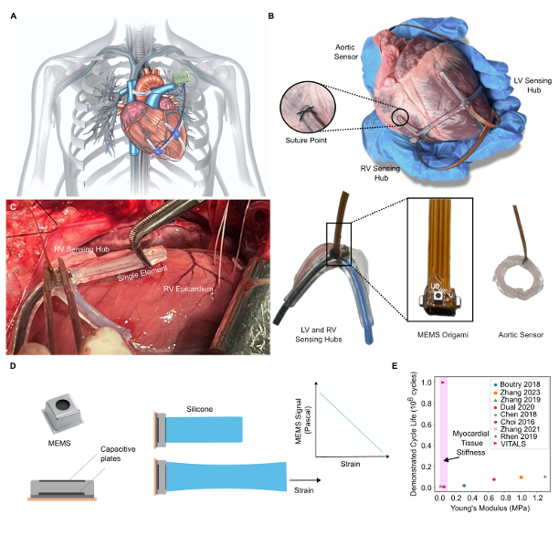
Figure 1. Illustration of the VITALS network and associated electronics implanted on a patient's epicardium B) Visualization of the VITALS network affixed to an ex-vivo porcine heart. C) Image of a single sensing hub attachment to the RV epicardium during an in vivo experiment. Mechanical energy transmission and transduction elements. B, C) Assembled sensor. D) A visualization of the sensing mechanism, whereby a capacitive micro-electromechanical system (MEMS) barometric pressure sensor is coupled to a soft silicone element.Sensor on the heart for GLS measurement E) comparison of VITALS to other stretchable strain sensors reported in literature with respect to demonstrated cycle life and stiffness..
Stage of Development
The inventors completed acute preclinical studies in a large animal model. They are currently integrating wireless capabilities.
Applications
- Post-operative cardiac monitoring
- Valve replacement
- Left ventricular assist device implantation
- Heart transplantation
- Coronary artery bypass grafting
- Cardiovascular drug and therapy research
Advantages
- Biocompatible
- Robust and durable
- Small enough to be implanted
- Uses same sensing mechanism as clinically deployed sensors (CardioMEMS), lowering developmental and clinical risk as well as making it well-suited for similar battery-less interrogation
- Highly stretchable and low stiffness
- Continuous monitoring
- Objective measure of cardiac health
- Experimentally validated to provide clinically relevant measurements such as preload and stroke volume, global longitudinal strain (GLS), and peak systolic pressure
Publications
- Kight, A., Pirozzi, I., et al. (2023). Decoupling Transmission and Transduction for Improved Durability of Highly Stretchable, Soft Strain Sensing: Applications in Human Health Monitoring. Sensors (Basel, Switzerland), 23(4), 1955.
Related Links
Patents
- Published Application: 20240263933
- Issued: 12,618,660 (USA)
Similar Technologies
-
Enhancing Heart Transplants with an Affordable "Any Size Fits All" Novel Ex-Vivo Perfusion System S24-135Enhancing Heart Transplants with an Affordable "Any Size Fits All" Novel Ex-Vivo Perfusion System
-
Primate Model and Magnesium Clock Mechanism for Cardiac Pacemaker Research and Arrhythmia Drug Discovery S25-149Primate Model and Magnesium Clock Mechanism for Cardiac Pacemaker Research and Arrhythmia Drug Discovery
-
Convex Formulation of Continuous Time Heartbeat Dynamics S23-288Convex Formulation of Continuous Time Heartbeat Dynamics