Docket #: S19-161
New powerful electron microscopy imaging mode combining Raman spectroscopy and electron microscopy
Stanford researchers at the Dionne Lab have introduced new vibrational spectroscopy, termed electron- and light induced stimulated Raman (ELISR) scattering, in electron microscopy for simultaneous high-resolution chemical mapping of various samples. This new powerful electron microscopy imaging mode enables Raman spectral imaging with spatial resolution approaching that of the electron-beam and simultaneous light and electron beam imaging for perfect imaging correlation. In addition, this imaging mode can be easily integrated with currently available transmission electron microscopes (TEMs), scanning electron microscopes (SEMs) and other instruments with electron beam imaging capabilities. Currently, there is no other technique that offers Raman spectral imaging in a TEM or SEM with the spatial resolution approaching that of the electron beam.
This new imaging mode can be used for label-free molecular and chemical mapping of biological and solid-state samples. It can also be used to characterize optical and chemical processes at the nanoscale for photocatalysis and energy storage process among other crucial applications.
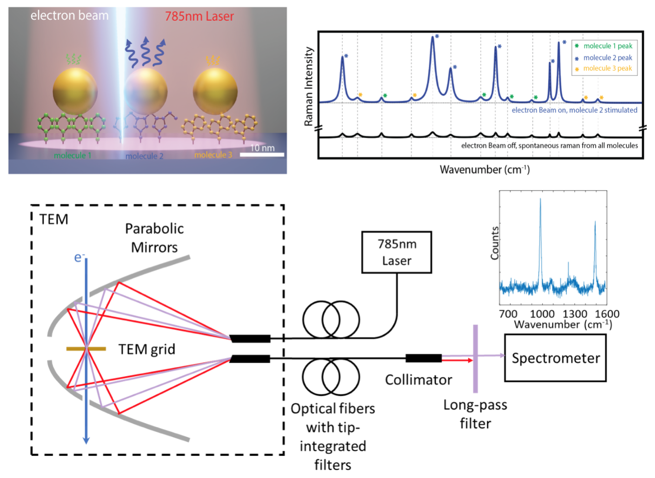
Figure Description - The top panel shows a schematic of ELISR. In the absence of the electron beam excitation, the Raman scattering from all illuminated molecules contributes to the spectra, prohibiting spatial localization below the diffraction limit (black illustrative spectrum). When the electron beam excites the plasmonic mode of one of the nanoparticles, the Raman scattering from the surrounding molecules is significantly enhanced while the scattering from the other molecules around unexcited nanoparticle remain unchanged (blue illustrative curve). Therefore, the molecular composition of a sample can be mapped with nanometer resolution. Experimentally, the team uses an aberration corrected environmental transmission electron microscope (TEM) with optical coupling ability. Two parabolic mirrors are used to illuminate the sample with the laser pump and collected the Raman scattering through two optical fibers under simultaneous e-beam excitation.
Applications
- Mapping of cells (cancer cells, bacteria, neurons…etc.) and other biological samples
- Characterize the local chemical composition of a variety of samples during electron imaging, including two-dimensional materials, polymeric blends and semiconductor devices.
- Study of photocatalysis and chemical reactions at the nanoscale
Advantages
- New features:
- Sub-wavelength Raman imaging with combined optical and electron excitation
- Simultaneous electron and stimulated Raman spectral imaging
- High chemical sensitivity with atomic spatial resolution approaching that of the electron-beam
- Easy implementation:
- Can be integrated with current commercial systems
- No specific labeling
- Same sample preparation for electron microscopy imaging
Publications
- Saleh, Amr AE, Daniel K. Angell, and Jennifer A. Dionne Electron and Light Induced Stimulated Raman Spectroscopy for Nanoscale Molecular Mapping" Preprint (2020).
- Dionne, Jennifer A., Fariah Hayee, Michal Vadai, Daniel Angell, Amr Saleh, and Katherine Sytwu. "The Light Years: Combined optical and environmental electron microscopy to visualize photonic processes with atomic-scale resolution." Microscopy and Microanalysis 25, no. S2 (2019): 2064-2065.
Similar Technologies
-
Multi-pass electron microscopy for enhanced imaging S17-348Multi-pass electron microscopy for enhanced imaging
-
Photoabsorption microscopy using electron analysis S18-070Photoabsorption microscopy using electron analysis
-
Multi-pass microscopy for high sensitivity, low damage microscopy S15-188Multi-pass microscopy for high sensitivity, low damage microscopy