Docket #: S19-233
Retrospective Tuning of MR Image Contrast for Precision Imaging
Stanford researchers at the Xing Lab have developed a novel technique to enable retrospective tuning of soft tissue contrast in MRI (i.e. adjusting the contrast after the image acquisition) using a deep learning-based strategy.
From just a single MR image, more tissue contrasts can be obtained, enabling full exploitation of MRI contrast versatility without additional cost.
In contrast, the conventional method to change tissue contrast is to acquire every new image using different imaging parameter values that are predetermined prior to data acquisition. More recently, MRI contrast can be retrospectively tuned based on quantitative parametric maps that are acquired using a special pulse sequence. In these cases, an additional data acquisition is required, which is costly and time consuming. The invention we propose has the promise to adapt tissue contrast in MRI without any additional data acquisition, providing optimal and to personalized contrast for precision imaging.
Related Technology:
Stanford docket S18-437 "Simultaneous acquisition of Qualitative and Quantitative MRI (Q2MRI) using deep learning". (Q2MRI) can derive quantitative relaxation parametric maps from single qualitative images. This principle is applied to this current invention.
Figure
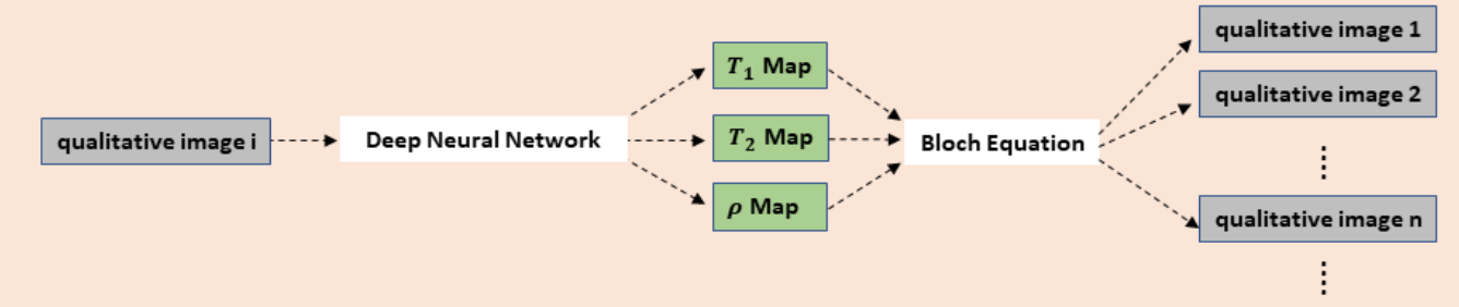
Figure description - Scheme of retrospective tuning of tissue contrast in MRI: from a single image, tissue relaxation parametric maps are derived and used to calculate images presumably acquired using other imaging protocols, providing a wide spectrum of tissue contrasts.
Stage of Development
Prototype
Applications
- Optimize soft tissue contrast of MR images for individual patients such that pathology (e.g. tumors) can be distinguished from surrounding normal tissues
Advantages
- Novel - retrospective tuning of MR image contrast from a single conventional MR image has never been achieved previously
- High accuracy - the anatomic structure and tissue contrast of generated images have high fidelity to the ground truth
- Provides additional information at no additional cost
- Only one conventional MR image acquired in a standard clinical scan is needed
- A wide spectrum of tissue contrast can be generated
- Provides optimal visualization of pathology for individual patients
- Generic framework that permits diversity in both input and output images
- Example application of Stanford docket S18-437
Publications
- Yan Wu, Yajun Ma, Jiang Du, and Lei Xing "Deciphering tissue relaxation parameters from a single MR image using deep learning", Proc. SPIE 11314, Medical Imaging 2020: Computer-Aided Diagnosis, 113140Q (16 March 2020)
- Wu, Yan, et al. "Quantitative Parametric Mapping of Tissues Properties from Standard Magnetic Resonance Imaging Enabled by Deep Learning." arXiv preprint arXiv:2108.04912 (2021).
Related Links
Patents
- Published Application: 2021-034706
- Issued: 11,675,029 (USA)
Similar Technologies
-
Simultaneous acquisition of Qualitative and Quantitative MRI (Q2MRI) using deep learning S18-437Simultaneous acquisition of Qualitative and Quantitative MRI (Q2MRI) using deep learning
-
Configurations for integrated MRI-linear accelerators S08-282Configurations for integrated MRI-linear accelerators
-
Radiotransparent audio-visual system to avoid pediatric patient anesthesia during radiation therapy and imaging. S23-306Radiotransparent audio-visual system to avoid pediatric patient anesthesia during radiation therapy and imaging.