Docket #: S21-387
Optical clearing enabled by the Kramers-Kronig relation
Researchers in the Brongersma and Hong labs have developed a novel method for optical clearing of turbid biological tissues.
Studying how cells behave within the 3-dimensional context of the biological structures they make up is critical for fully understanding tissue and organ function. While optical imaging techniques are fundamental tools of biological and medical research, they are intrinsically limited to studying extremely thin sections of biological tissues due to how these tissues scatter light. The primary cause of this light scattering is the heterogeneous mix of refractive indeces within biological tissues. Several techniques have been developed to correct this refractive index mismatch by replacing low-index water with high-index organics or vice versa; however, these techniques often involve toxic reagents and cannot be used in live tissues.
This new optical clearing method from the Brongersma and Hong labs leverages a distinct physical phenomenon compared to existing methods, features low-toxicity, and can even be used for optical clearing of live in vivo tissues and organs.
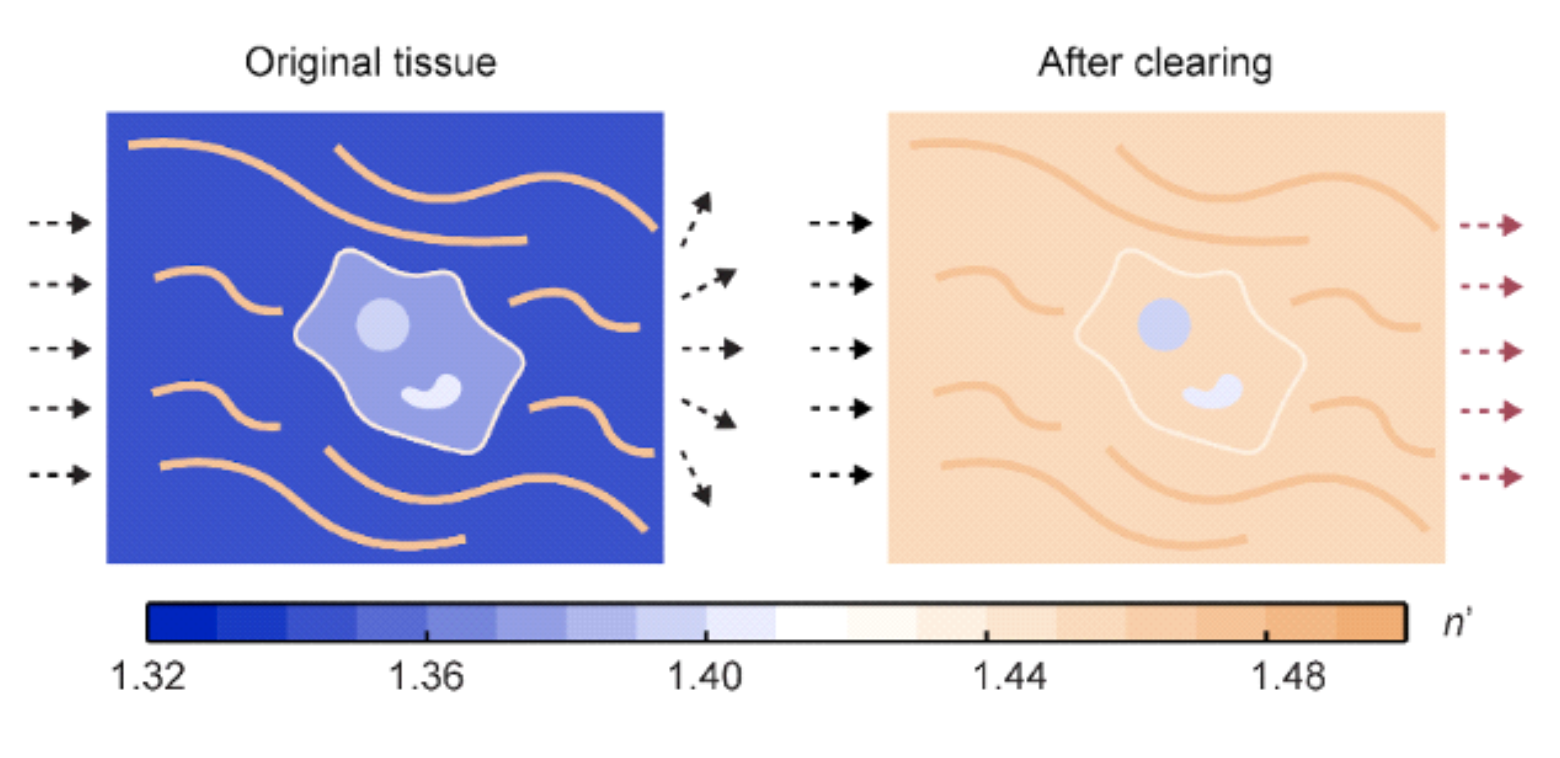
Fig. 1
Applications
- Optical/Tissue clearing of turbid biological tissues
- Optical/Tissue clearing of live tissues
- Optical/Tissue clearing of in vivo tissues
Advantages
- Low-toxicity
- applicable to live tissues
- applicable to in vivo tissues and organs
Publications
- Zihao Ou et al. Achieving optical transparency in live animals with absorbing molecules. Science , 385 (2024).
- Miller, Jen A. Researchers make mouse skin transparent using a common food dye. Stanford Report Sep 2024.
- A common food dye can make skin transparent. The Economist Sep 2024.
Related Links
Similar Technologies
-
Light sheet fluorescence microscopy using high speed structured and pivoting illumination S16-332Light sheet fluorescence microscopy using high speed structured and pivoting illumination
-
Multi-Functional In Vivo Cardiovascular Imaging Using Near-Infrared II Fluorescence S12-195Multi-Functional In Vivo Cardiovascular Imaging Using Near-Infrared II Fluorescence
-
Ultra Bright Lanthanide-Doped Nanoparticles for Luminescence Imaging S18-147Ultra Bright Lanthanide-Doped Nanoparticles for Luminescence Imaging