Docket #: S18-332
Rapid axial scanning light sheet microscope
Stanford researchers designed and built a light sheet microscope that can be used for deconvolution-free, high resolution volumetric imaging of cleared tissue specimens. The Solgaard lab microscope design uses a grating light valve (GLV) to scan the focus of a gaussian beam, creating a thin light sheet with the linear phased array acting as a varifocal cylindrical lens (Figure 1). During the scan the narrowest region of the beam (the line focus) is synchronized with the center of a rolling camera shutter that captures the image. The camera only captures the image from the narrowest part of the beam, thereby significantly increases axial resolution. The design includes a high-speed phase modulator, so the beam scan maintain pace with the camera (the system is camera limited), and isotropic resolution is possible with a narrow focus. This light sheet microscope maintains real-time, volumetric imaging capabilities beyond current state of the art.
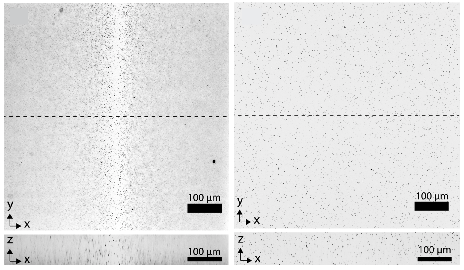
Fig. 1 (Inverted image) Light sheet nominal focus with the phased array off (left) and uniformly illuminated light sheet (right) with synchronization of rolling shutter and beam focus as designed.
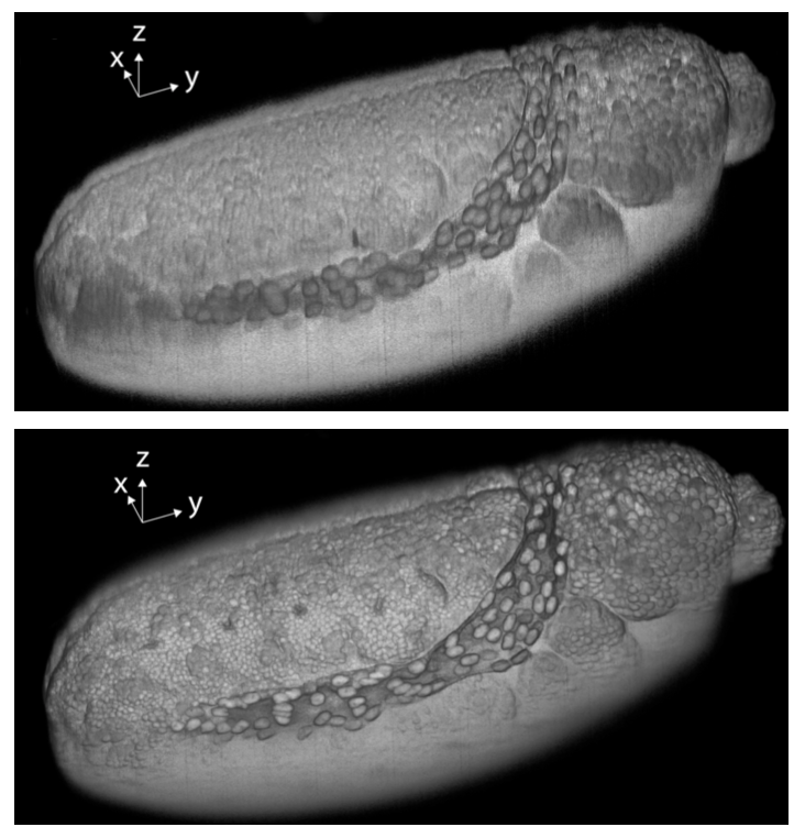
Fig. 2 Drosophila embryo 3D images showing the surface of the reconstructed 3D volumes with rolling shutter widths of 1625 (top) and 65 ?m (bottom), respectively.
Stage of Development – Proof of Concept Prototype
Researchers in the Solgaard Lab built and tested the axial scanning light sheet microscope. Initial design is fast and simple, but there are limits to the maximum obtainable isotropic resolution. Future research includes testing an improved GLV with smaller pixels to scan narrower spot sizes across the field-of-view and simultaneously correct for aberrations caused by scanning along the optical axis of high Numerical Aperture objectives. Smaller pixels will also enable lateral scanning, which facilitates 3D volume imaging without moving the sample.
Applications
- Light Sheet Microscopy
- Optical Coherence Tomography
- LiDAR
- Replacement for confocal microscope
Advantages
- Fast, real-time, high resolution 3D imaging.
- Free from time consuming 3D deconvolution.
- Aberration free over large sample FOV.
- Simple design that is easy to automate and incorporate autofocus.
Publications
- Landry, Joseph, Stephen Hamann, and Olav Solgaard. "High-speed axially swept light sheet microscopy using a linear MEMS phased array for isotropic resolution." Journal of Biomedical Optics 25, no. 10 (2020): 106504. https://doi.org/10.1117/1.JBO.25.10.106504
Patents
- Published Application: 20200371032
- Issued: 11,619,585 (USA)
Similar Technologies
-
Method and Apparatus for Evaluating Electrostatic or Nonlinear Devices S19-023Method and Apparatus for Evaluating Electrostatic or Nonlinear Devices
-
Breakthrough Optical Frequency Processing for Quantum Computing and Beyond S24-365Breakthrough Optical Frequency Processing for Quantum Computing and Beyond
-
Efficient wide-field nanosecond imaging methods using Pockels cells for low-light applications S18-388Efficient wide-field nanosecond imaging methods using Pockels cells for low-light applications